Publications
Magnify
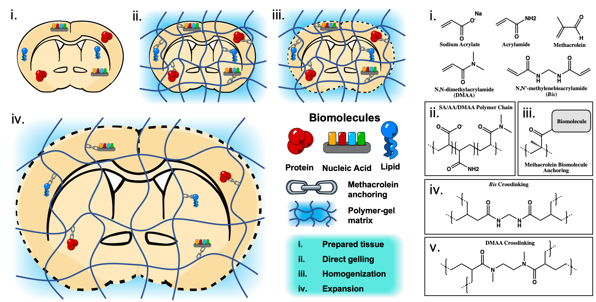
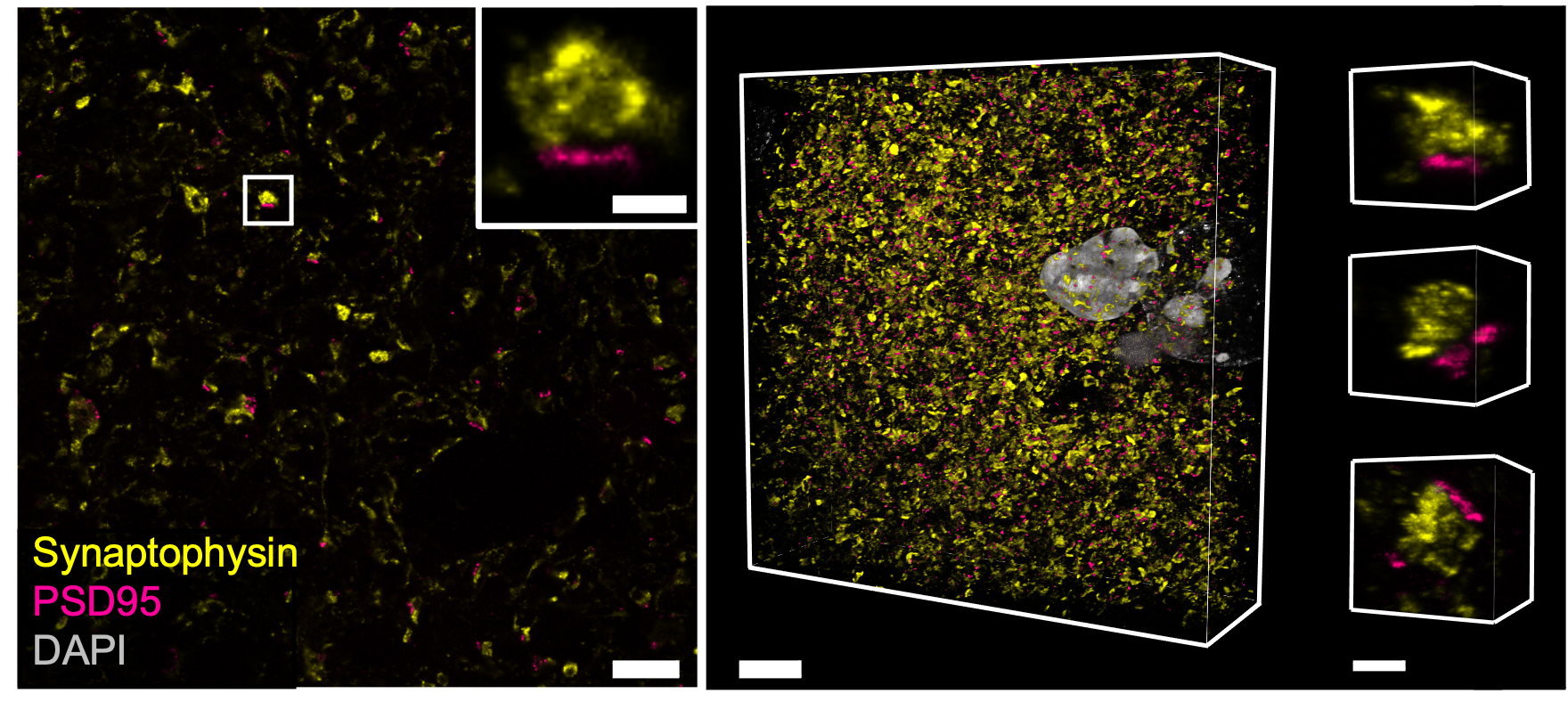
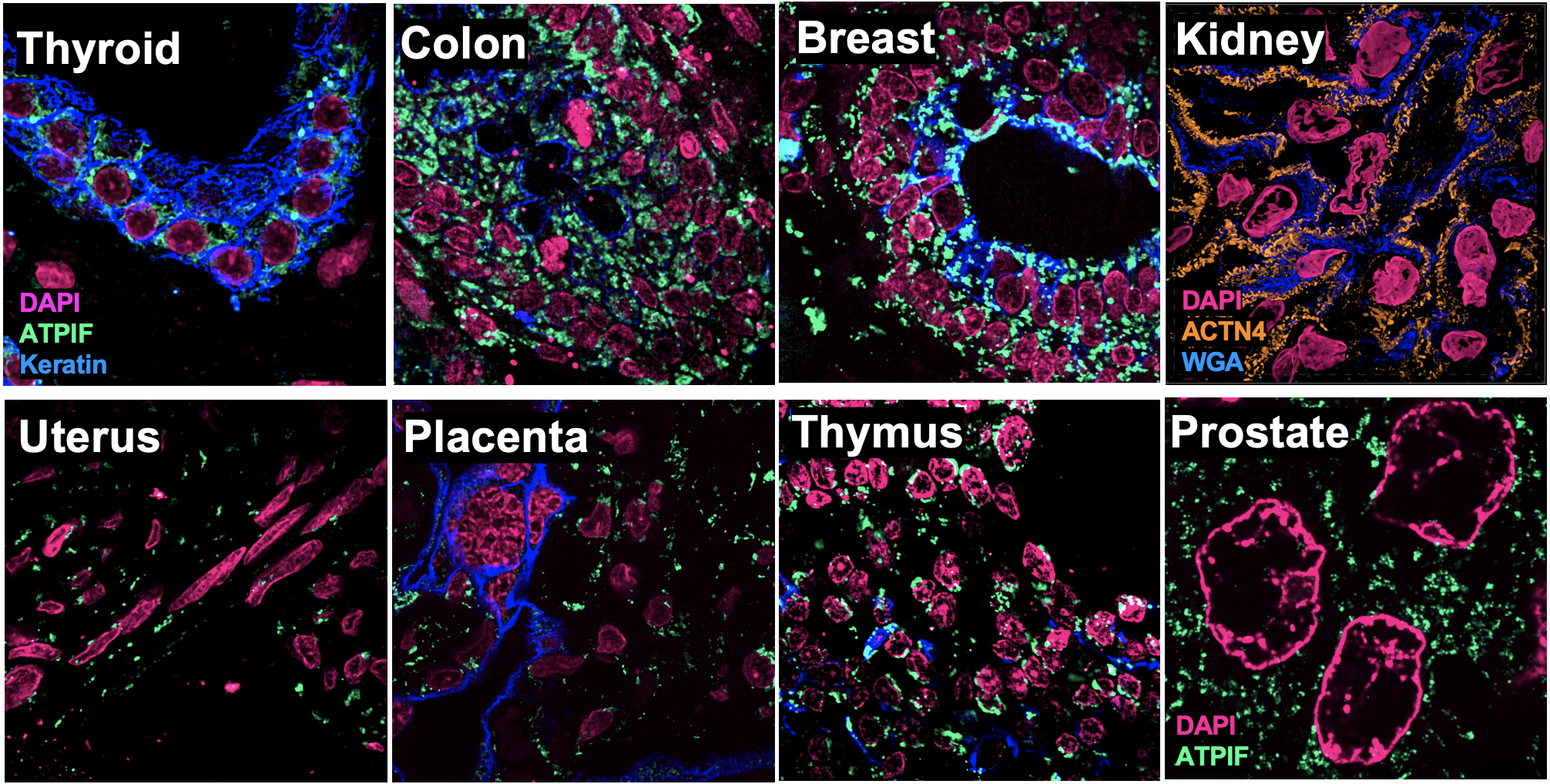
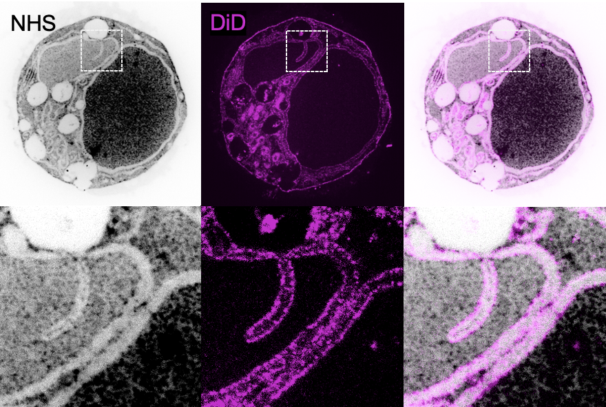
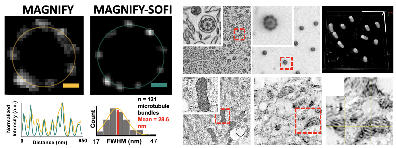
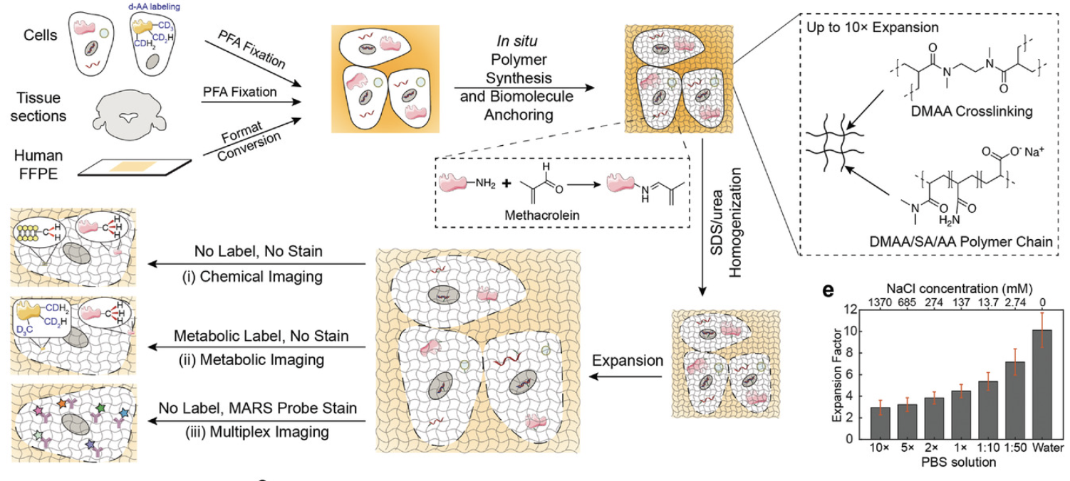
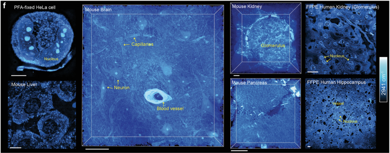
Abstract: Expansion microscopy (ExM) enables nanoimaging with conventional microscopes by physically and isotropically magnifying preserved biological specimens embedded in a cross-linked water-swellable hydrogel. Current ExM protocols require prior treatment with reactive anchoring chemicals to link specific labels and biomolecule classes to the gel. We describe a new strategy called Magnify, which uses a mechanically sturdy gel that retains nucleic acids, proteins, and lipids without the need for a separate anchoring step. Magnify expands biological specimens up to 11× and facilitates imaging of cells and tissues with effectively ~25-nm-resolution using a ∼280-nm diffraction-limited objective lens on conventional optical microscopes or with ~15 nm effective resolution if combined with Super-resolution Optical Fluctuation Imaging (SOFI). We demonstrate Magnify on a broad range of biological specimens, providing insight into nanoscopic subcellular structures including synaptic proteins from mouse brain, podocyte foot processes in formalin-fixed paraffin-embedded human kidney, and defects in cilia and basal bodies in drug-treated human lung organoids.
Klimas, A., Gallagher, B.R., Wijesekara, P. et al. Magnify is a universal molecular anchoring strategy for expansion microscopy. Nat Biotechnol (2023).
MAGNIFIERS
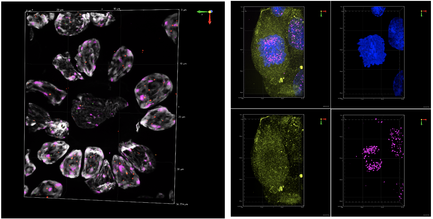
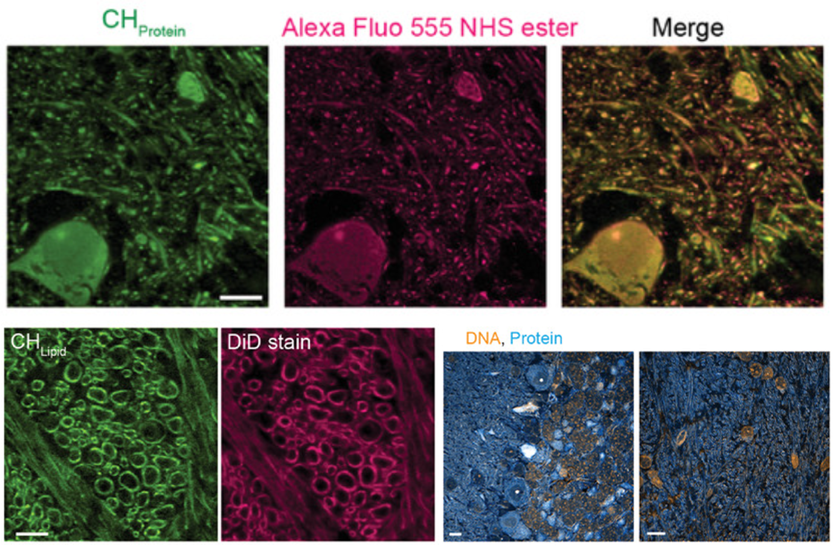
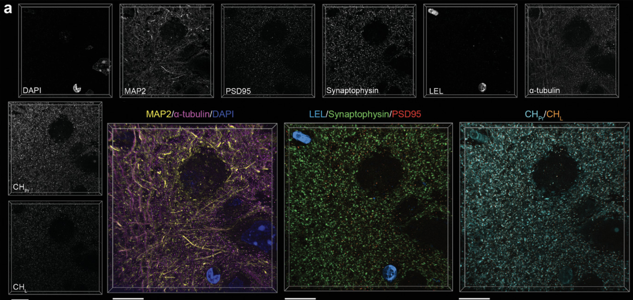
Abstract: Stimulated Raman scattering (SRS) microscopy is an emerging technology that provides high chemical specificity for endogenous biomolecules and can circumvent common constraints of fluorescence microscopy including limited capabilities to probe small biomolecules and difficulty resolving many colors simultaneously. However, the resolution of SRS microscopy remains governed by the diffraction limit. To overcome this, a new technique called molecule anchorable gel-enabled nanoscale Imaging of Fluorescence and stimulated Raman scattering microscopy (MAGNIFIERS) that integrates SRS microscopy with expansion microscopy (ExM) is described. MAGNIFIERS offers chemical-specific nanoscale imaging with sub-50 nm resolution and has scalable multiplexity when combined with multiplex Raman probes and fluorescent labels. MAGNIFIERS is used to visualize nanoscale features in a label-free manner with C-H vibration of proteins, lipids, and DNA in a broad range of biological specimens, from mouse brain, liver, and kidney to human lung organoid. In addition, MAGNIFIERS is applied to track nanoscale features of protein synthesis in protein aggregates using metabolic labeling of small metabolites. Finally, MAGNIFIERS is used to demonstrate 8-color nanoscale imaging in an expanded mouse brain section. Overall, MAGNIFIERS is a valuable platform for super-resolution label-free chemical imaging, high-resolution metabolic imaging, and highly multiplexed nanoscale imaging, thus bringing SRS to nanoscopy.
, , , , , , , , , , Super-Resolution Vibrational Imaging Using Expansion Stimulated Raman Scattering Microscopy. Adv. Sci. 2022, 9, 2200315. https://doi.org/10.1002/advs.202200315